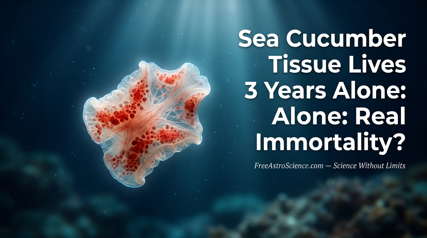
Sea Cucumber Tissue Lives For Three Years Without a Body: A Discovery That Rewrites Biology
What if a small piece of an animal could heal, grow, and stay alive for years after being cut away from its body, with no sterile lab, no nutrients, no special care? Welcome to FreeAstroScience.com, friends. We’re glad you’re here. Today we’re walking you through a finding that genuinely surprised us, and we promise it’ll be worth your time. Stick with us to the end. The story we’re about to tell challenges what biologists have believed for two centuries.
📑 What you’ll find inside
- ➤ A discovery that breaks the rulebook
- ➤ Who is Psolus fabricii?
- ➤ What happens in week one?
- ➤ How can a body part eat without a mouth?
- ➤ From day six to year three
- ➤ Why this species and no other?
- ➤ Why does this change medicine?
- ➤ FAQ
A Discovery That Breaks the Rulebook {#discovery}
For 200 years, biologists have tried to keep animal tissue alive outside its host body . The results were modest. Embryonic and larval cells could be cultured, sure, but only inside sealed flasks with carefully prepared nutrient media . Adult tissues? They decayed within days or weeks. Period.
Then came the team led by Sara Jobson at Memorial University of Newfoundland. Working with Annie Mercier, Jean-François Hamel, Rachel Sipler, and Emaline Montgomery, they cut small pieces of tissue from a cold-water sea cucumber called Psolus fabricii and dropped them into running, non-sterile seawater .
What happened next belongs in a textbook. The pieces healed. They grew back. They kept living for more than three years.
The researchers gave these tissues a name: LiPfe, short for Living Immortal Psolus fabricii Explants. The work appeared in Science Advances on May 27, 2026.
Who Is Psolus fabricii? {#species}
This isn’t a glamorous animal. Psolus fabricii is a small dendrochirotid sea cucumber found across the Atlantic and Arctic at depths of 8 to 13 meters . Its sole sticks to rocks. Its branching tentacles fan out to catch food drifting by .
It belongs to the phylum Echinodermata, alongside starfish and sea urchins. Here’s the part that matters: echinoderms are deuterostomes, like us. They share a deep evolutionary link with vertebrates, which makes their biology more relevant to human medicine than you might think .
Echinoderms are already famous for two things:
- Strong regenerative powers (sea stars regrow arms)
- Negligible cellular senescence — their cells barely show signs of aging
But what P. fabricii did pushes well beyond regeneration. The pieces didn’t just survive on a parent body. They survived alone.
What Happens In Week One? {#week-one}
The team excised tube feet, ambulacra (groups of tube feet), tentacles, and body wall sections from three adult sea cucumbers . They placed each fragment in small culture wells covered with mesh, with natural seawater flowing through. No antibiotics. No sterile cabinet. No enriched broth .
Here’s the early timeline.
| Day | What the researchers saw |
|---|---|
| Day 0 | Damaged tissue at wound margin; immune cells already active |
| Day 1 | Peak mitosis (57.61 m²s⁻²) and apoptosis (39.03 m²s⁻²) |
| Day 1–2 | Necrotic tissue shed; healthy edges curl inward |
| Day 3 | Second peak of cell division and programmed cell death |
| Day 6 | 100% of explants showed full wound closure |
| Diameter | Shrunk 23%, from 2.10 mm to 1.59 mm during initial healing |
The healing followed a clear, almost choreographed sequence: clean, reorganize, regenerate, maintain . Two opposite forces drove it forward, oscillating roughly every 24 hours. Mitosis built new cells. Apoptosis cleared damaged ones. They worked in tandem, not at random .
The role of immune cells
Sea cucumbers carry their own internal defense force called coelomocytes . These cells migrated straight to the wound after cutting. At day 0, the epidermal tissue carried up to 5,367 coelomocytes per mm². By day 6, the count had dropped 54% as the cleanup phase wound down . In the connective tissue, the drop reached 84% .
The immune system, in short, was already doing its job. Without help. Without instructions.
How Can a Body Part Eat Without a Mouth? {#feeding}
This question stumped us at first. A tube foot has no stomach. So how does it survive for three years?
The team designed a clever isotope test. They exposed explants to ¹⁵N-labeled algal amino acids (a mix of 16) or ¹⁵N-labeled ammonium chloride, then measured how much the tissue absorbed .
The result: after 6 days, amino acid absorption climbed from 0.37 atom% (control) to 1.34 atom% in treated explants — a statistically significant increase (t₁₈ = 7.74, P < 0.001) .
Translation: the tissue absorbed dissolved nutrients straight from seawater through its outer surface . It didn’t need a digestive tract. The ocean fed it.
There was a second twist. Muscle tissue inside the explant, with no purpose without a body to move, slowly disappeared. The researchers watched coelomocytes phagocytize it, eating muscle for nutrients . By six months, no muscle remained . The tissue cannibalized itself to keep living.
From Day Six to Year Three {#year-three}
Here’s the long-term story.
| Time | Observation |
|---|---|
| 30–60 DPE | Wound site becomes invisible — fully merged with surrounding tissue |
| 60–120 DPE | Explants regrow to original size |
| 122 DPE | Color shifts; pigmented cells migrate inward |
| 180 DPE | Explants now 21% larger than original |
| 365 DPE | Connective tissue takes over (74% of explant); pigments form one large red mass |
| 730 DPE (2 yr) | Same morphology preserved; transparent connective tissue around periphery |
| 912 DPE (≈2.5 yr) | Explants “remained largely unchanged” |
| >3 years | Some explants survived buried under ~10 mm of mud, alongside other organisms |
The tentacles kept moving
This blew our minds. Tentacle explants didn’t just survive. They kept responding to touch, extending and retracting after tactile stimulation . Synaptic transmission was still working. A piece of nervous tissue, alive and reactive, with no brain attached .
A 1928 study once reported a chick embryo heart that kept beating in culture . But those embryonic tissues never responded to their environment. The Psolus fabricii tentacles did .
Why This Species and No Other? {#unique}
The team didn’t stop at P. fabricii. They tested tissue from six other echinoderms :
- Hippasteria phrygiana (sea star): 104 DPE
- Strongylocentrotus droebachiensis (sea urchin): 74 DPE
- Leptasterias polaris (sea star) & Cucumaria frondosa (sea cucumber): ~55 DPE
- Ophiopholis aculeata (brittle star): ~55 DPE
- Chiridota leavis: 42 DPE
- Body wall samples (all species): <42 DPE
Only Psolus fabricii showed indefinite survival . Why?
The researchers point to psolusosides — triterpene glycosides unique to this species, with documented antimicrobial and cytotoxic properties . They might give P. fabricii a built-in chemical shield that keeps fouling bacteria at bay .
It’s a working hypothesis. Future studies will need to confirm it. But the contrast with the other six species is striking.
Why Does This Change Medicine? {#why-matters}
Tissue culture has leaned for decades on two pillars: HeLa cells, the famous immortal human cervical cancer line discovered in the 1950s, and stem cells from mice, identified in the 1960s . Both reproduce indefinitely in vitro. Neither produces an organized tissue with multiple coordinated cell types .
LiPfe close that gap.
What makes this model different?
- No ethical barriers — invertebrate tissue, not human or vertebrate
- No sterile equipment — runs in plain natural seawater
- Low cost — accessible to schools and small labs
- Multiple coordinated tissue types — epidermal, connective, muscle, neural
- Real-time platform for testing pollutants, drugs, and xenobiotics
- Wound healing studies with possible insights for human skin regeneration
The authors put it bluntly: this challenges “conventional perceptions of tissue immortality” . It’s a new class of experimental model. One that bridges evolutionary biology with translational medical research .
We’re not saying human skin will start healing for three years in saltwater. That’s not the point. The point is: nature has solved a problem we’ve struggled with for 200 years. Studying how could open doors we haven’t even mapped yet.
A Note Before We Close
We wrote this article specifically for you on FreeAstroScience.com, where we explain complex science in simple words. We do this because we believe you should never switch off your mind. As Goya warned us, the sleep of reason breeds monsters. Keep questioning. Keep reading. Keep your curiosity alive.
Closing Thoughts
A small piece of sea cucumber tissue, no bigger than a pea, healed itself, fed itself from seawater, defended itself with its own immune cells, and stayed alive for more than three years in plain running seawater . It even survived buried under mud, surrounded by other organisms .
This isn’t a footnote in biology. It rewrites the line between “alive” and “dying.” It hands medicine a model with no ethical baggage, no sterile lab requirement, and stunning resilience . And it reminds us that the deep ocean still holds answers we haven’t dreamed of asking about.
Come back to FreeAstroScience.com soon. We’ll keep digging into discoveries that matter, and we’ll keep your mind moving.
FAQ {#faq}
Q1: What does LiPfe mean?
LiPfe stands for Living Immortal Psolus fabricii Explants — pieces of sea cucumber tissue (tube feet, tentacles, ambulacra) that survived more than three years in natural, non-sterile seawater .
Q2: How long did the tissue actually live?
The formal experiment ran for one year. Opportunistic monitoring extended observations beyond 3 years (over 1,095 days), with explants still showing tissue maintenance .
Q3: Why didn’t the tissue rot?
Three reasons: (1) coelomocytes — the sea cucumber’s immune cells — neutralized bacteria around the wound; (2) the tissue absorbed dissolved amino acids from seawater for energy; (3) P. fabricii likely produces psolusosides, antimicrobial compounds that prevent fouling .
Q4: Could this work directly on human tissue?
No, not directly. Human tissue lacks coelomocytes and these specific antimicrobial compounds. But studying LiPfe could reveal mechanisms that inspire new therapies for wound healing, skin regeneration, and infection-resistant tissue engineering .
Q5: Where and when was the study published?
In Science Advances, volume 12, article eaeb1394, on May 27, 2026, by Sara Jobson and colleagues from Memorial University of Newfoundland, Vancouver Island University, the SEVE Society, and the Bigelow Laboratory for Ocean Sciences .
📚 References
- Jobson, S., Montgomery, E. M., Hamel, J.-F., Sipler, R. E., & Mercier, A. (2026). Natural tissue immortality: Indefinite survival of sea cucumber explants. Science Advances, 12, eaeb1394. science.org