
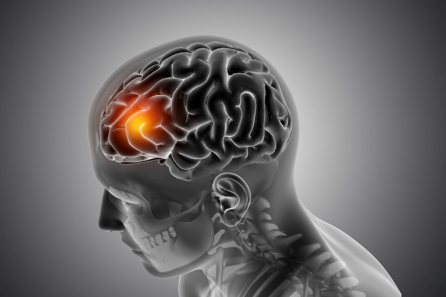
Brain aneurysms are small bulges in the vessels of the brain that can rupture without warning and lead to strokes. About one US adult in 50 harbors this insidious threat, but doctors remain unable to work out which ones are most lethal.
Now, a new study from UC San Francisco offers a breakthrough glimpse into the biology behind these events, showing how certain cells inside the brain’s arteries may drive aneurysms toward rupture. The findings, published June 10 in Nature Neuroscience, could reshape how doctors think about risk and open doors to new ways of preventing strokes before they happen.
The UCSF team analyzed more than 100,000 individual cells from human aneurysms and healthy brain arteries. What they uncovered was a cast of 19 distinct cell types, each playing a role in the vessel wall’s health or decline.
Normally, arteries are composed of three organized layers: an inner layer (endothelium), a middle layer of smooth muscle cells, and an outer layer of fibroblasts. This structure ruptures during aneurysms. Degraded and replaced by scar formers called “activated fibroblasts” that stiffen the vessel up and express genes associated with aneurysm risk instead of smooth muscle cells.
“We’ve made major steps toward solving the mystery of how aneurysms form,” said Ethan Winkler, MD, PhD, assistant professor of Neurological Surgery and senior author of the study. “We’ve identified the cast of characters involved and seen which ones are implicated at different phases of disease.”
Scientists discovered a destructive feedback loop in which arterial fibroblasts induce macrophages to secrete enzymes that erode blood vessels. Blocking those signals stopped the macrophages from producing the damaging enzymes.
In the following selective phase, weakening of the annulus, disappearance of muscle cells, dense sustained scar tissue that hardens the wall itself, and immune cells promote faster destruction. This is why small aneurysms are still dangerous, as Winkler pointed out: more than 50% of ruptures happen below the magic surgical boundary of 7 millimeters.
“We’ve had to rely on anatomy because we haven’t known the underlying biology,” Winkler said.
By uncovering the cellular drivers of rupture, the research suggests that size alone is not enough to predict danger. Biology matters, and now, scientists are beginning to decode it.
State-of-the-art treatment of aneurysms is size- and location-specific and also accounts for patient risk factors. These are biologically stabilized by blocking fibroblast signals or avoiding other immune recognition.
“Maybe one day we’ll be able to stabilize an aneurysm to prevent it from bursting,” Winkler said. “That would be a very effective treatment, and one we’ve dreamed of for a long time.”
This study reframes aneurysms not just as structural flaws but as dynamic ecosystems where cells interact in harmful ways. By mapping those interactions, scientists are moving closer to predicting which aneurysms are most dangerous and, perhaps one day, preventing rupture altogether.
For patients and doctors alike, that could mean turning a silent threat into a manageable condition and rewriting the story of one of medicine’s most feared strokes.